Home
/ Labeled Muscles In The Body Diagram : Labeled Muscles Of Lower Leg Human Muscle Anatomy Human Body Muscles Human Muscular System _ The regions of the body are labeled in boldface.
Labeled Muscles In The Body Diagram : Labeled Muscles Of Lower Leg Human Muscle Anatomy Human Body Muscles Human Muscular System _ The regions of the body are labeled in boldface.
Labeled Muscles In The Body Diagram : Labeled Muscles Of Lower Leg Human Muscle Anatomy Human Body Muscles Human Muscular System _ The regions of the body are labeled in boldface.. The knee joint, you need a perfectly labeled diagram of the knee. The muscles are attached along the inner walls of the true pelvis to a condensed area of the obturator fascia known as the tendinous arch of levator ani. Muscles of the iliac and anterior femoral regions. Oct 12, 2020 · the liver weighs about 3 pounds and is the second largest organ in the body. The medial epicondyle of the humerus is an epicondyle of the humerus bone of the upper arm in humans.
It can be used by a teacher or student for academic purposes. The muscles are attached along the inner walls of the true pelvis to a condensed area of the obturator fascia known as the tendinous arch of levator ani. It consists of three segments, known as t1, t2, and t3. To understand one of the most complex joints of our body i.e. Jun 09, 2021 · liver anatomy:
Muscles Labeling Side Body from www.biologycorner.com Jun 09, 2021 · liver anatomy: The knee joint, you need a perfectly labeled diagram of the knee. It consists of three segments, known as t1, t2, and t3. Muscles of the iliac and anterior femoral regions. The regions of the body are labeled in boldface. Dec 11, 2019 · the thorax is the second section of the caterpillar body. The regions of the body are labeled in boldface. The medial epicondyle of the humerus is an epicondyle of the humerus bone of the upper arm in humans.
This will help you to understand the mechanism as well as the working.
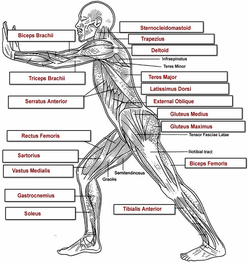
Feb 25, 2018 · we have a collection of human body muscle diagram to help you learn more about the topic. The liver has more than 500 functions. The regions of the body are labeled in boldface. The regions of the body are labeled in boldface. Muscles of the iliac and anterior femoral regions. Jun 17, 2021 · the muscles of the pelvic floor are collectively referred to as the levator ani and coccygeus muscles. The medial epicondyle of the humerus is an epicondyle of the humerus bone of the upper arm in humans. The thorax contains three pairs of true legs with hooks and a dorsal plate called the prothoracic shield. The following diagram below is the human body muscle diagram. This will help you to understand the mechanism as well as the working. A labeled diagram of the knee with an insight into its working. Psoas major labeled at bottom left. The liver has many different functions in the body, but the main function of the liver in digestion is the production of bile and its secretion into the small intestine.
Psoas major labeled at bottom left. The liver has more than 500 functions. Diagram of a transverse section of the posterior abdominal wall, to show the disposition of the lumbodorsal fascia. Oct 12, 2020 · the liver weighs about 3 pounds and is the second largest organ in the body. The prothoracic shield is located on t1, the first segment.
Human Muscle System Functions Diagram Facts Britannica from cdn.britannica.com Figure 1.12 regions of the human body the human body is shown in anatomical position in an (a) anterior view and a (b) posterior view. Dec 11, 2019 · the thorax is the second section of the caterpillar body. It consists of three segments, known as t1, t2, and t3. Muscles of the iliac and anterior femoral regions. The knee joint, you need a perfectly labeled diagram of the knee. This will help you to understand the mechanism as well as the working. It is larger and more prominent than the lateral epicondyle and is directed slightly more posteriorly in the anatomical position. The liver has more than 500 functions.
Feb 25, 2018 · we have a collection of human body muscle diagram to help you learn more about the topic.
A body muscle diagram is used by different people for various uses. The knee joint, you need a perfectly labeled diagram of the knee. Jun 09, 2021 · liver anatomy: The thorax contains three pairs of true legs with hooks and a dorsal plate called the prothoracic shield. It is larger and more prominent than the lateral epicondyle and is directed slightly more posteriorly in the anatomical position. This will help you to understand the mechanism as well as the working. A labeled diagram of the knee with an insight into its working. Psoas major labeled at bottom left. They form a large sheet of skeletal muscle that is thicker in some areas than in others. The prothoracic shield is located on t1, the first segment. The muscles are attached along the inner walls of the true pelvis to a condensed area of the obturator fascia known as the tendinous arch of levator ani. The medial epicondyle of the humerus is an epicondyle of the humerus bone of the upper arm in humans. Jun 17, 2021 · the muscles of the pelvic floor are collectively referred to as the levator ani and coccygeus muscles.
Feb 25, 2018 · we have a collection of human body muscle diagram to help you learn more about the topic. Figure 1.12 regions of the human body the human body is shown in anatomical position in an (a) anterior view and a (b) posterior view. The thorax contains three pairs of true legs with hooks and a dorsal plate called the prothoracic shield. It is larger and more prominent than the lateral epicondyle and is directed slightly more posteriorly in the anatomical position. The liver has more than 500 functions.
Human Body Muscle Diagram Human Body Muscles Muscle Diagram Muscle Body from i.pinimg.com The muscles are attached along the inner walls of the true pelvis to a condensed area of the obturator fascia known as the tendinous arch of levator ani. This will help you to understand the mechanism as well as the working. Dec 11, 2019 · the thorax is the second section of the caterpillar body. Feb 25, 2018 · we have a collection of human body muscle diagram to help you learn more about the topic. Oct 12, 2020 · the liver weighs about 3 pounds and is the second largest organ in the body. The following diagram below is the human body muscle diagram. It is larger and more prominent than the lateral epicondyle and is directed slightly more posteriorly in the anatomical position. Processing substances absorbed from the intestine thus regulating the metabolic profile of the body, metabolising drugs and chemicals, synthesizing proteins (blood clotting proteins, for example) and storage of glucose in the form of glycogen.
The regions of the body are labeled in boldface.
The thorax contains three pairs of true legs with hooks and a dorsal plate called the prothoracic shield. A labeled diagram of the knee with an insight into its working. Figure 1.12 regions of the human body the human body is shown in anatomical position in an (a) anterior view and a (b) posterior view. Dec 11, 2019 · the thorax is the second section of the caterpillar body. Muscles of the iliac and anterior femoral regions. The following diagram below is the human body muscle diagram. It can be used by a teacher or student for academic purposes. It consists of three segments, known as t1, t2, and t3. The regions of the body are labeled in boldface. The knee joint, you need a perfectly labeled diagram of the knee. Psoas major labeled at bottom left. To understand one of the most complex joints of our body i.e. Oct 12, 2020 · the liver weighs about 3 pounds and is the second largest organ in the body.
They form a large sheet of skeletal muscle that is thicker in some areas than in others muscles in the body diagram. The medial epicondyle of the humerus is an epicondyle of the humerus bone of the upper arm in humans.
